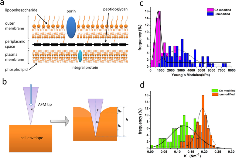
Figure 3.
a) Structure of the E. coli bacterial cell envelope showing the outer membrane, the peptidoglycan, and the inner plasma cell membrane. b) Distortion of the E. coli cell wall with a AFM tip indenting inward. c) Distribution of the Young’s modulus of the E. coli DH5α grown on CA-coated and uncoated PDMS surfaces. d) Distribution of the bacterial spring constant K of the E. coli DH5α growing on CA-coated and uncoated PDMS surfaces.

