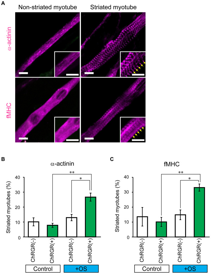
Figure 3. Effects of OS training on sarcomere assembly in ChRGR-expressing C2C12 myotubes.
(A) Images of typical multinucleated myotubes 7 days differentiation induction. Cells were fixed and stained with anti-sarcomeric α-actinin (upper panel) or fMHC (lower panel). The insets show the magnified images. The sarcomeric structures are indicated by arrows. Scale bar, 10 μm. (B) Fractions of myotubes with mature striation patterns. Assembly of α-actinin (from left to right): control ChRGR-negative myotubes without OS training (white column, n = 12 regions), ChRGR-positive myotubes without OS training (green column, n = 12 regions), ChRGR-negative myotubes with OS training (white column, n = 16 regions), and ChRGR-positive myotubes with OS training (green column, n = 16 regions). (C) Similar to (B), but showing the assembly of fMHC in ChRGR-negative (white bar, each, n = 16 regions) and –positive cells (green bar, each, n = 16 regions). Each experiment consists of at least three independent replicates. *, P < 0.05; **, P < 0.01; Kruskal–Wallis test.

