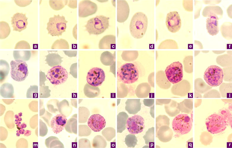
Fig. 1.

Microphotographs of Plasmodium ovale in Giemsa-stained thin blood films collected in Singapore. a–c ring stages, d, e young trophozoites, f trophozoite, g late trophozoite, h young schizont, i–k growing schizont, l late schizont, m ruptured schizont, n young gametocyte, o, p developing macrogametocytes, q macrogametocyte, r microgametocyte. a–e, h–m, o, q and r are P. ovale curtisi while f, g, n and p are P. ovale wallikeri
