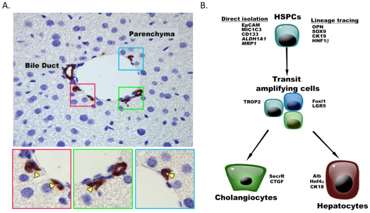
Figure 1. The niche of HSPCs and their expression markers .
(A) HSPCs are located in the smallest and most peripheral branches of the biliary tree, the ductules and canals of Hering. In healthy murine livers, canals of Hering are present in the portal area and consist of one hepatocyte and several cholangiocytes/HSPCs. By immunohistochemistry, cholangiocytes/HSPCs are identified by their Krt19-positivity (brown). Corresponding channels and their lumens are magnified in the lower panels (colored squares, yellow arrowheads indicate the lumen). (B) HSPCs can be isolated directly using FACS or MACS with antibodies directed to EpCAM, MIC1C3, CD133 or using a functional assay (ALDH1A1 (aldehyde activity), MRP1 (Side Population). When studying their functionality, lineage tracing of quiescent HSPCs is often used based on markers such as OPN, SOX9, CK19, HNF1β. Once activated, HSPCs become transit amplifying cells expressing the surface marker (TROP2) as well as Foxl1 and LGR5 which are also used for lineage tracing.

