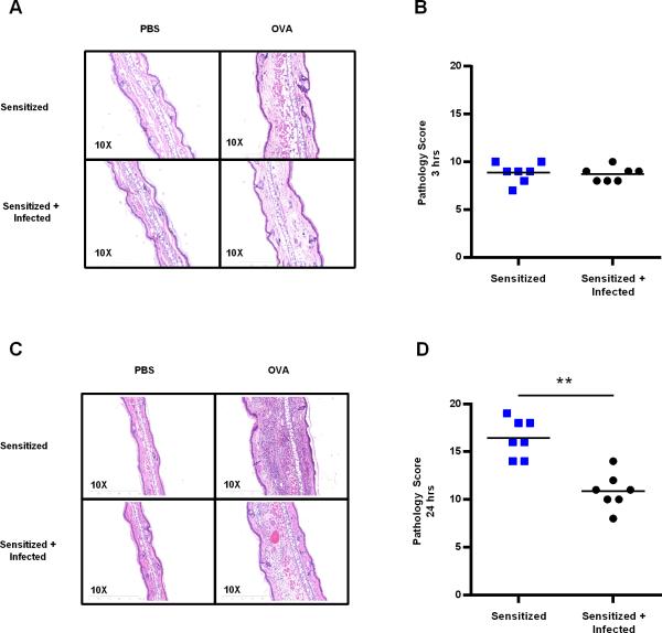
Figure 5.
Sensitized + Infected animals exhibited reduced ear pathology 24 hours post-challenge. (A) H&E stain of ear tissue at 3 hours post-challenge, the time point at which immune complexes were detected by immunohistochemistry. (B) Pathology score for 3 hour time point. Four parameters were measured (hemorrhaging, edema, necrosis, and cellularity) on the basis of severity (0-absent, 1-mild, 2-moderate, or 3-severe) and focality (0-absent, 1-focal, 2-intermediate, and 3-diffuse) for a total maximum score of 24. (C) H&E stain of ear tissue 24 hours post-challenge, the time point at which there was the greatest difference in ear swelling between groups. (D) Pathology score for 24 hour time point. Data are representative of two independent experiments with 3-4 BALB/c mice per group. Statistical differences between groups were analyzed by the Mann Whitney test. ** p < 0.01.

