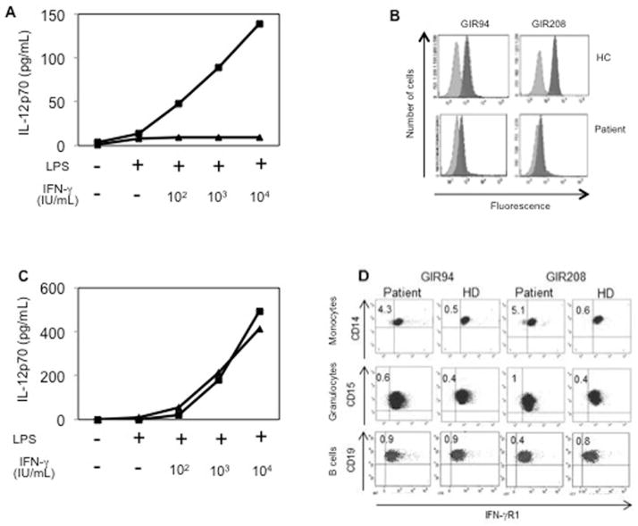
Figure 2. Cytokine responses to IFN-γ stimulation and expression of IFN-γR1 prior and after HSCT.
(A) IL-12p70 production in vitro in response to lipopolysaccharide plus various concentrations of IFN-γ before HSCT. The patient is represented by triangles and a healthy control (HC) with squares.
(B) Expression of IFN-γR1 before HSCT. Whole blood from the patient and from one HC was stained with two IFN-γR1-specific mAbs (dark gray; GIR94 and GIR208) and isotypic control antibodies (pale gray).
(C) IL-12p70 production in vitro response to lipopolysaccharide plus various concentrations of IFN-γ after HSCT as described in (A).
(D) Expression of IFN-γR1 after HSCT. Whole blood from the patient and from a HC was stained with two IFN-γR1-specific mAbs as done in (B). CD14+ monocytes, CD15+ granulocytes and CD19+ B cells were analyzed.

