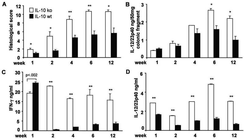
Figure 3. Time course of histological evaluation of colitis and proinflammatory cytokine production in recipient mice.
(A) Histologic score, (B) spontaneous IL-12/23p40 in colonic cultures, and amounts of (C) IFN-γ and (D) IL-12/23p40 produced by unfractionated MLN cells stimulated with CBL (10 μg/ml) after 3 day culture in vitro for mice evaluated 1, 2, 4, 6 and 12 weeks after transfer of SPF IL-10 ko CD4+ T cells to IL-10 ko Rag2−/− or IL-10 wt Rag2−/− recipients. ** p< 0.001 and *p<0.05 or p value shown above the bar vs. IL-10 wt Rag2−/− recipients. Results show mean ± SEM, 6-8 mice per group at each time point.

