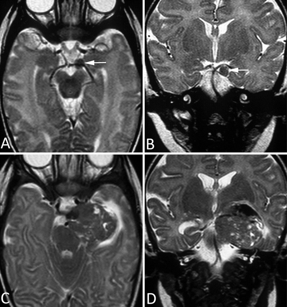
Fig 3. MRI Images of Patient 3.
A–B. Initial MRI at presentation. a. Axial postcontrast T1-weighted image shows a solid, avidly enhancing lesion at the CN III origin, anterior to the right cerebral peduncle. b. Axial ADC image shows iso- to hypo-intense signal within the mass and moderately increased signal within adjacent brainstem parenchyma, indicating edema.
C–D. Follow-up MRI 1 week after presentation. c. Axial postcontrast T1-weighted image and (d.) precontrast ADC image show slight interval growth of the mass despite the very short follow-up interval.
E–F. Follow-up MRI 1 month after presentation. e. Axial postcontrast T1-weighted image and (f.) precontrast ADC image clearly demonstrate interval growth of the lesion, with two new remote foci of abnormal contrast enhancement—one in the right ambient cistern (arrowhead) and one in the olfactory region in the medline (arrowhead)—suggestive of leptomeningeal dissemination.
G–H. Follow-up MRI 5 weeks after presentation. g. Axial postcontrast T1-weighted image and (h.) precontrast ADC image show further interval growth of both the primary lesion and the metastatic deposit.

