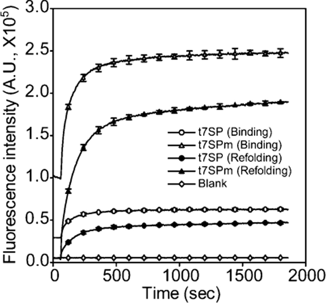
Figure 5.
Time-dependent fluorescence recovery of the leave-one-out GFP. The fluorescence recovery of the leave-one-out GFP upon in vitro complementation is shown. Chemically synthesized peptides (s7) with a purity of >75% were used. The complementation was initiated by adding peptide solutions (pH 7.5) to equilibrated native t7SP and t7SPm (white symbols) or by bringing the pH of protein/ peptide mixtures from 2.0 to 7.5 (black symbols). Final concentrations of the leave-one-out GFP (t7SP and t7SPm) and s7 in both experiments were 0.1 and 3.6 µM, respectively. The fluorescence intensity at 508 nm emission upon the complementation was recorded and plotted every 5 s for 30 min with excitation at 485 nm at room temperature. Error bars indicate the two standard deviations from triplicate measurements and were provided only every 120 s.

