Figure 2.
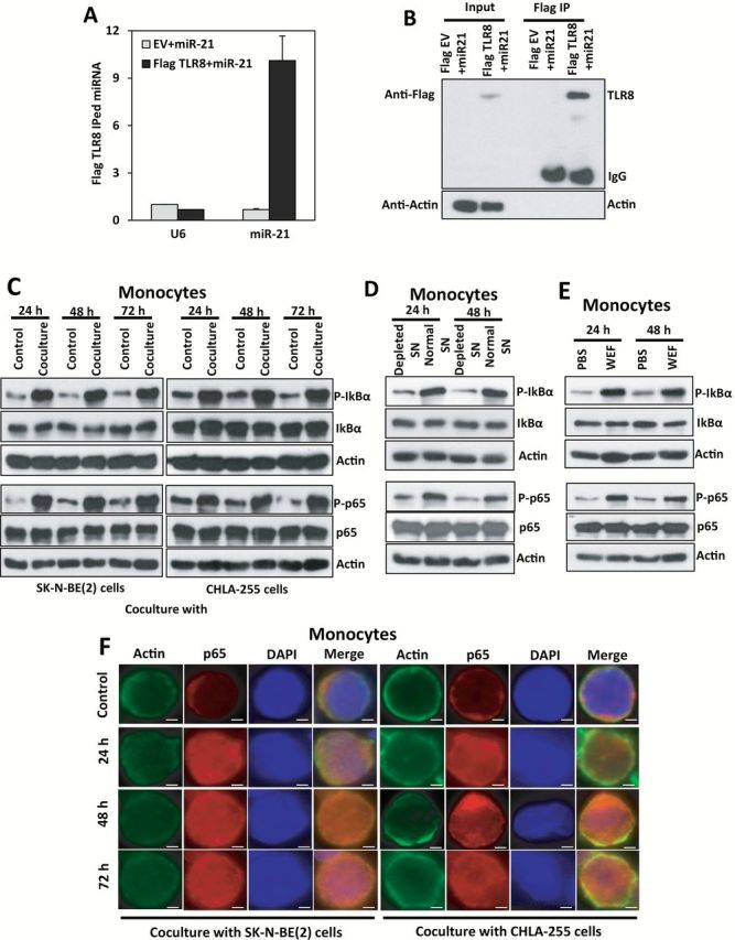
Exosomic miR-21 and TLR8 interaction in human monocytes. A) Quantitative real-time polymerase chain reaction (qRT-PCR) for miR-21 or U6 in the immunoprecipitate with anti-Flag antibody in human monocytes transfected with empty vector and Dotap-miR-21 (EV+miR-21) or with a Flag-TLR8 vector and Dotap-miR-21 (Flag TLR8+miR-21), after 48 hours from trasfection. Data presented as mean ± SD of experiments conducted in triplicate. B) Immunoblotting with anti-Flag antibody corresponding to the experiment described in (A). C) Immunoblotting for phospho-IкBα, IкBα, phospho-p65, p65, and relative actin proteins in human monocytes cocultured with SK-N-BE(2) or CHLA-255 cells for the indicated time periods. D) Immunoblotting for phospho-IкBα, IкBα, phospho-p65, p65, and relative actin proteins in human monocytes cultured with the supernatant (SN) from SK-N-BE(2) cells that were depleted for exosomes (depleted SN) compared with normal SN for the indicated time periods.
(E) Immunoblotting for phospho-IкBα, IкBα, phospho-p65, p65 and relative actin proteins in human monocytes treated with whole exosomes fraction (WEF) isolated from SK-N-BE(2) cells compared with PBS for the indicated time periods. F) Immunofluorescence (IF) image showing intracellular localization of the p65 NF-кB subunit in human monocytes cocultured with SK-N-BE(2) or CHLA-255 cells. IF staining was performed using monoclonal anti-p65 (red) and polyclonal anti-actin (red) antibodies together with DAPI (blue). Results are a representative of three independent experiments. Scale bar = 2 μm.