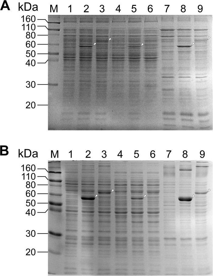
FIG 5.
Expression of phcC and phcD in Sphingobium sp. SYK-6 and E. coli. Proteins (10 μg) were separated on SDS-12% polyacrylamide gels and stained with Coomassie brilliant blue. (A) Lanes: 1, 4, and 7, SYK-6 harboring pJB861 (vector); 2, 5, and 8, SYK-6 harboring pJBI09480 (phcC); 3, 6, and 9, SYK-6 harboring pJBI09500 (phcD); 1 to 3, cell extracts; 4 to 6, soluble fractions; 7 to 9, membrane fractions. (B) Lanes: 1, 4, and 7, E. coli BL21(DE3) harboring pET-16b (vector); 2, 5, and 8, E. coli BL21(DE3) harboring pET09480 (phcC); 3, 6, and 9, E. coli BL21(DE3) harboring pET09500 (phcD), 1 to 3, cell extracts; 4 to 6, soluble fractions; 7 to 9, membrane fractions; M, molecular mass markers.

