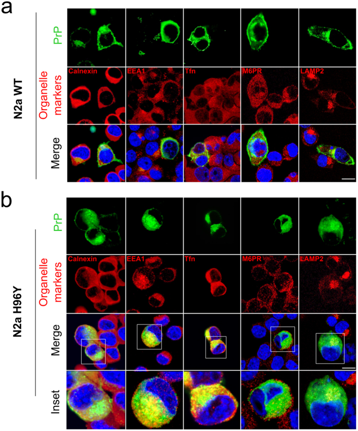
Figure 6. The H96Y MoPrP mutant displays intracellular accumulation in the endosomial compartments.
PrP localization in N2a cells expressing the 3F4-WT MoPrP (a) or H96Y MoPrP (b). Nuclei are labeled with DAPI (blue), PrPs are detected by 3F4 antibody (green); organelle markers, such as Calnexin (ER marker), EEA1 (early endosomes marker), Tfn (recycling endosome marker), M6PR (late endosome marker) and LAMP2 (lysosome marker) are labeled in red. Insets in (b) shows a magnification of the merged panels (white boxed areas). Scale bars: 12 μm.

