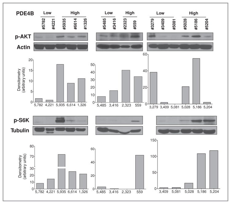
Figure 5.
AKT/mTOR activity correlates with PDE4B expression in primary DLBCL. Western blot analyses of phospho-AKT (S473) and phospho-S6K (T389) were done in primary DLBCLs categorized by PDE4B expression (see Supplementary Table S3). Densitometric analysis, normalized by 2 independent proteins (β-actin and α-tubulin), is also shown and points to a correlation between PDE4B expression and activity of the AKT/mTOR pathway in the majority of primary DLBCLs analyzed and a significantly higher expression of these phospho-proteins in PDE4B-high DLCBLs (P < 0.05, Mann–Whitney test for the densitometric values). Note that protein from sample 3279 was available for only one of the Western blot analysis.

