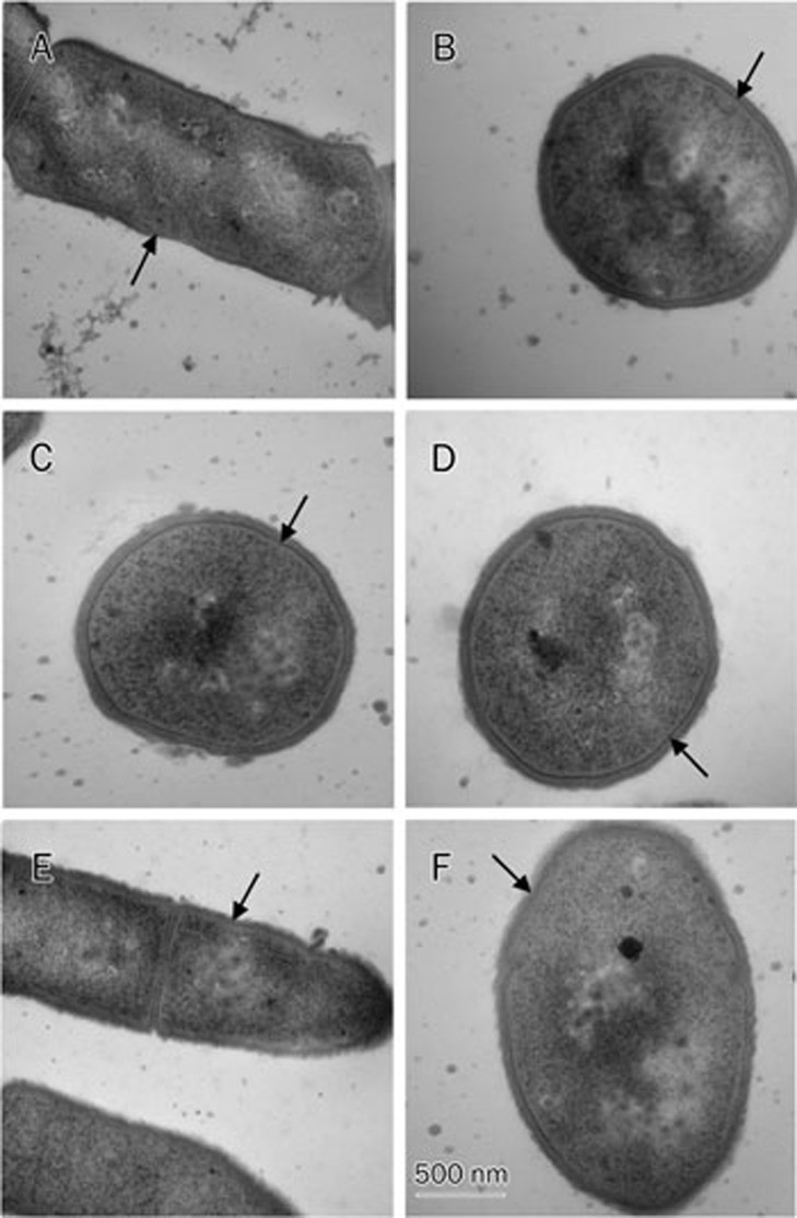
Figure 6.
Transmission electron micrographs of B anthracis AP422 cells treated with daptomycin and other antibiotics. (A) and (B) MHBc control; (C) 1 μg/mL daptomycin for 30 min; (D) 4 μg/mL daptomycin for 30 min; (E) 1 μg/mL penicillin G for 30 min; and (F) 25 μg/mL nisin for 30 min. No significant structural damage to the cytoplasmic membrane of B anthracisAP422 cells that were treated with daptomycin was observed. Arrows point to the cell membrane; Scale bar, 500 nm.

