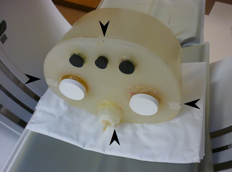
Figure 2.
Photograph showing the phantom positioned on the CT table in the gantry. Visible on the sides of the phantom are the markings used to ensure alignment (arrowheads). Also visible are the kidney and spine inserts, as well as three plugged holes that can accommodate simulated gastric tubing (not used in this study).

