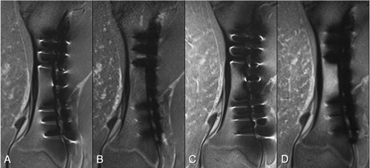
Figure 5.
Tissue specimen containing one plate and six stainless steel screws evaluated at 1.5 T (a, b) and 3.0 T (c, d), with proton density-weighted conventional sequence (a, c) and slice encoding for metal artefact correction with view angle tilting (SEMAC-VAT) technique (b, d). At both field strengths a substantial reduction in metal artefact was provided by SEMAC-VAT, allowing for better visualization of the tissue structures adjacent to the metal implants.

