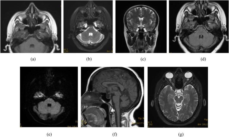
Figure 1.
A 38-year-old female presented with headache. Axial T1 weighted (a), axial T2 weighted (b), coronal T2 weighted (c) and axial fluid-attenuated inversion recovery (d) MR images show bilateral petrous apex cephalocoele with cerebrospinal fluid content continuous with the Meckel's cave (arrows). Diffusion restriction is not noted on a diffusion-weighted image (e). Sagittal T1 weighted MR image shows coexistent empty sella (f). Axial T2 weighted MR image shows coexistent bilateral optic nerve sheath diameter distention (g).

