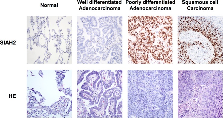
Fig 4. Correlation between SIAH2 expression and tumor grade.
Representative images of immunohistochemical analysis of SIAH2 in normal lung, well and poorly differentiated adenocarcinoma and squamous cell carcinoma. Occasional nuclear positivity in normal lung, moderate staining in well differentiated adenocarcinoma and strong staining in poorly differentiated adenocarcinoma and squamous cell carcinoma (HE: hematoxylin–eosin)(x100).

