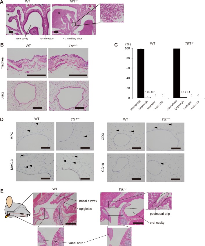
Fig 3. Postnasal drip detected in the pharynx of Ttll1 −/− mice without lower airway inflammation.
(A) Coronal sections of the nasal cavity and paranasal sinus stained with hematoxylin and eosin (HE). Accumulation of mucus and neutrophils were found in the nasal cavity and sinus of the Ttll1 −/− mice. Scale bar, 200 μm. (B) Trachea and lung sections stained with HE. Inflammation findings were not observed in the tracheae and lungs of the Ttll1 −/− mice. Scale bar, 200 μm. (C) Differential cell counts in bronchoalveolar lavage fluid obtained from the wild-type (WT) and Ttll1 −/− mice. There were no significant differences between the WT and Ttll1 −/− mice (mean ± SEM, n = 4 mice per group, two-paired Student's t test). (D) Lung sections stained for myeloperoxidase (MPO), Mac-3, CD3, and CD19. A few inflammatory cells (arrows) are detected in the peribronchial region. There were no significant differences between the WT and Ttll1 −/− mice. Scale bar, 200 μm. (E) Sagittal sections of the upper airway stained with HE. Postnasal drip characterized by accumulation of mucus and neutrophils was found in the pharyngeal wall. There was no inflammation in the larynx of the Ttll1 −/− mice. Scale bar, 500 μm.

