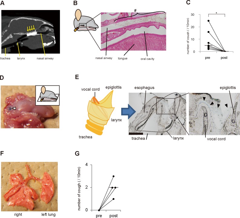
Fig 7. Laryngeal stimuli by postnasal drip-evoked cough in mice.
(A) Contrast material in the nasal airway and upper airway was scanned by computed tomography (CT). CT shows optimal nasal airway obstruction with contrast material (arrows). (B) Physical blockade of the nasal airway to dam postnasal drip. Cyanoacrylate glue was placed in the nasal airway of Ttll1 −/− mice, as illustrated. Sagittal section of the nasal airway stained with hematoxylin and eosin showing obstruction of the nasal airway with cyanoacrylate glue (#). (C) Graph displaying the pre- and post-treatment number of coughs in the Ttll1 −/− mice. Cough in the Ttll1 −/− mice was completely inhibited by nasal airway blockade with cyanoacrylate glue (bars: median values, n = 7, *P = 0.02 by Wilcoxon signed-rank test). (D−F) Artificial postnasal drip in the wild-type (WT) mice (n = 5). A blue-colored polyvinyl alcohol (PVAL) solution was intranasally administered to the WT mice to mimic postnasal drip. The photos show the lower jaw (D) and lung (F), and the photomicrographs show sagittal sections of the larynx (E) after administration of the PVAL solution. The PVAL solution (blue) was found in the larynx (white and black arrowheads). Bar, 1 mm. There was no finding of aspiration of the PVAL solution in the trachea and lungs (E and F). (G) Graph showing the pre- and post-treatment number of coughs in the WT mice. A PVAL solution (artificial postnasal drip) was intranasally administered to the WT mice. Cough was evoked by an artificial postnasal drip in the WT mice (n = 4).

