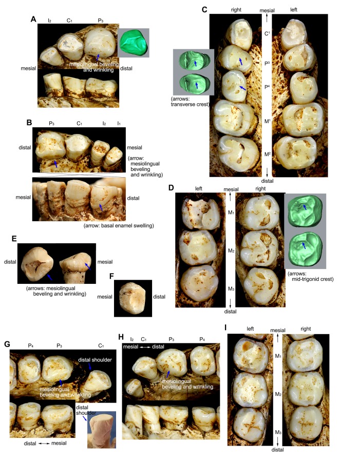
Fig 1. Teeth of Homo floresiensis.
Right (A) and left (B) anterior dentitions of the LB1 mandible. (C) Maxillary dentition of LB1 with EDJ surface images for the right P3 and P4. (D) Mandibular molars of LB1. (E) Occlusal (left) and lingual (right) views of the LB2/2 left P3. (F) Occlusal view of the LB15/1 right P4. Left (G) and right (H) anterior dentitions of the LB6/1 mandible with a photograph of a cast of its left C1 (with blue background). (I) Mandibular molars of LB6/1. See ref. [24] for LB 15/2 (I1) and LB6/14 (I1).

