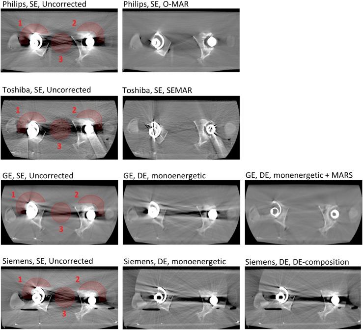
Figure 2.
CT images of the hip prostheses shown for the CT scanners from four different vendors: Philips Healthcare (Cleveland, OH), Toshiba Medical Systems (Otawara, Japan), GE Healthcare (Milwaukee, WI), and Siemens Healthcare (Forchheim, Germany) (for details, see Table 1). The first column shows the uncorrected images acquired with a tube voltage of 120 kVp with the three regions of interest marked. The second and third columns show the images obtained with the different metal artefact reduction (MAR) techniques (MAR algorithms and/or monoenergetic reconstructions of dual-energy (DE) CT data). All images shown are reconstructed with filtered backprojection and a soft reconstruction kernel. A window width of 1400 and a window level of 300 are used for displaying the images. MARS, metal artefact reduction software; O-MAR, MAR for orthopaedic implants; SE, single energy; SEMAR, single-energy MAR.

