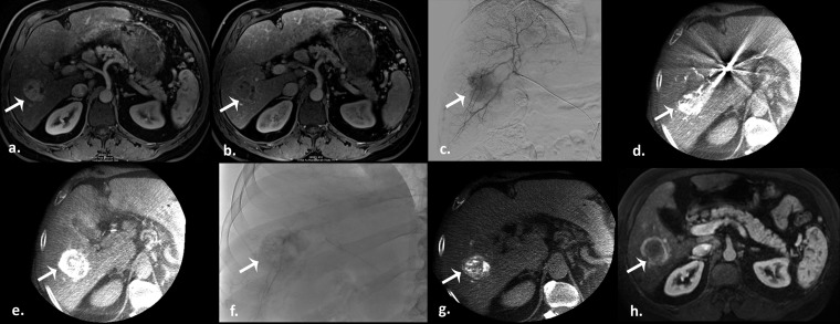
Figure 2.
Drug-eluting beads transarterial chemoembolization in a 55-year-old male with a history of nonalcoholic steatohepatitis. (a) Pretreatment arterial phase T1 weighted gradient-echo MR scan demonstrates a hyperenhancing hepatocellular carcinoma in the right hepatic lobe, with subsequent washout on the portal venous phase (b) (arrows). (c) Selective digital subtraction angiography clearly demonstrates tumour blush (arrow). Intraprocedural pretreatment dual-phase cone beam CT (CBCT) in early (d) and late (e) arterial phases evidence the lesion (arrows). Postembolization single-snap shot (f) and CBCT (g) show an excellent contrast staining of the tumour (arrows). (h) Arterial phase T1 weighted gradient-echo MR scan obtained 1 month after therapy demonstrate a good result (partial response) (arrow).

