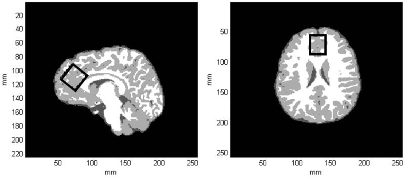
Fig. 1.
(a) A tissue-segmented axial slice extracted from a 3D MPRAGE dataset recorded from a 17-year-old female HC subject. WM, GM and CSF are represented by white, light gray and dark gray pixels, respectively. (b) A tissue-segmented sagital slice extracted from the same MPRAGE dataset. The black rectangle depicts the positioning of the MRS voxel within the ACC, which was obliqued along the sagital dimension. For this subject, GM, WM and CSF tissue fractions were estimated to be 74, 24 and 2%, respectively.

