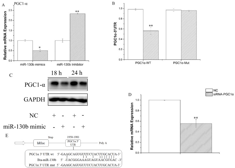
Fig 5. PGC-1α was identified as a target of miR-130b in GMECs.
A: GMECs were transfected with miR-130b mimic or inhibitor for 48h, PGC-1α expression level was quantified by RT-qPCR (n = 4). White bars, negative control; black bars, miR-130b mimic or inhibitor. B and E: Target site of miR-130b in PGC-1α 3’UTR and the construction of the luciferase (Luc) expression vector fused with the PGC-1α 3’UTR. WT, Luc reporter vector with the WT PGC-1α 3’UTR (1958 to 1981); MU, Luc reporter vector with the mutation at miR-130b site in PGC-1α 3’UTR. C: Western blot analysis of PGC-1α expression in the miR-130b mimic and NC treatment experiments. The effect of miR-130b mimics on PGC-1α protein expression was evaluated by western blot analysis in GMECs. Total protein was harvested 24 h or 18 h post-transfection, respectively. NC, negative control. D: GMECs were transfected with siRNA of PGC-1α for 48h later, miR-130b expression levels was quantified by RT-qPCR (n = 6). All experiments were performed in duplicate and repeated three times. Values are presented as means ± standard errors, *, P<0.05; **, P<0.01

