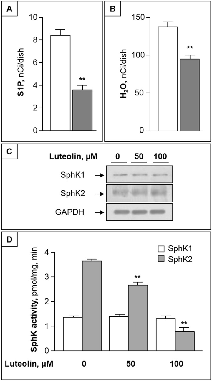
Fig 5. Luteolin reduces cellular S1P by inhibiting SphK2.
(A) and (B) CC cells were pulsed with 3H-Sph for 2 h in the absence (white bar) or presence of 100 μM luteolin (grey bar). At the end, labeled S1P (A) and water (B) were evaluated in cells and medium, respectively. (C) CC cells were treated with 50–100 μM luteolin for 2h, and equal amounts of proteins were then analyzed for SphK1 and SphK2 proteins by immunoblotting. GAPDH was used as loading control. (D) SphK1 and SphK2 activities were assayed in the absence or presence of luteolin, using CC cell homogenate as enzyme source. Data are mean ± S.D. of three experiments in duplicate. **, p < 0.01 vs. control.

