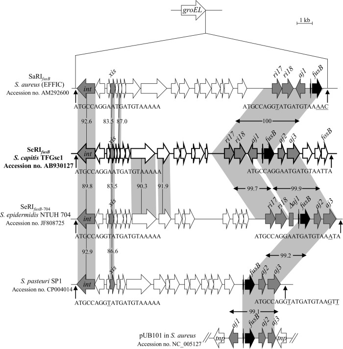
Fig 1. Structure of ScRIfusB in S. capitis subsp. urealyticus TFGsc1.
ScRIfusB was compared to SaRIfusB, SeRIfusB-704, the RI in the S. pasteuri genome (the above RIs are inserted into groEL) and the plasmid pUB101. The ORFs are shown as arrows, and the genes of interest are indicated as grey or black arrows. The homologous regions are shaded, and the numbers in the shadow show the percent homology between the corresponding sequences in comparison to ScRIfusB. The predicted att sites are indicated by vertical arrows. Th divergent nucleotides in the 21-bp att sequences are underlined.

