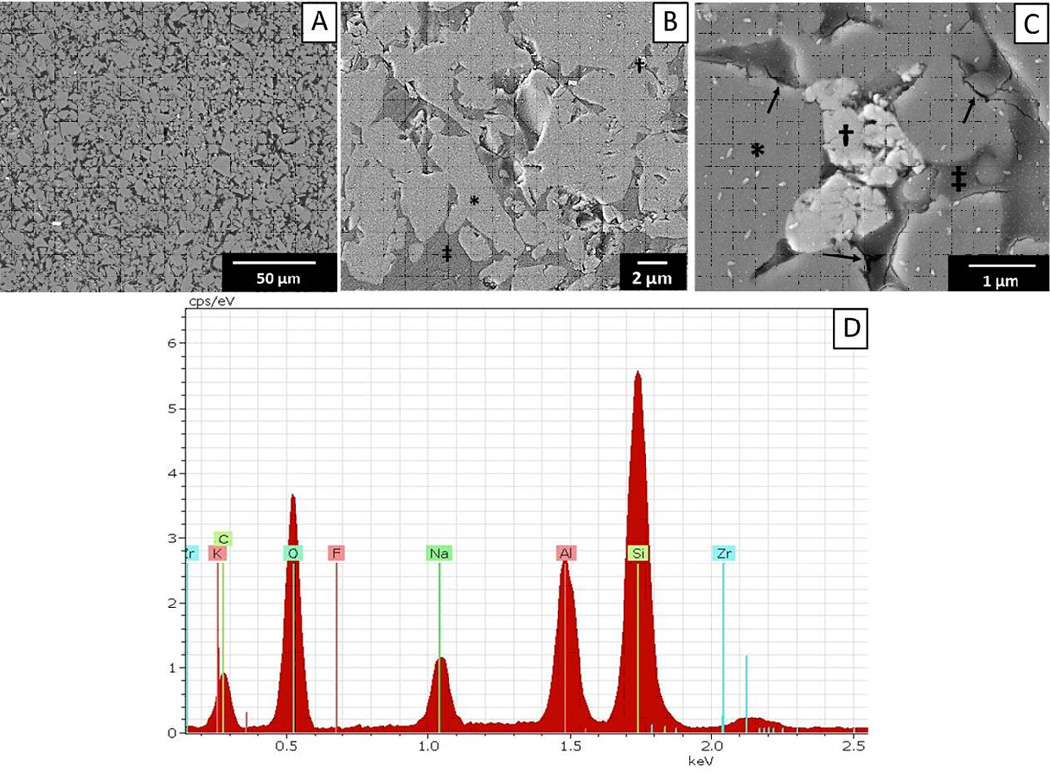
Figure 3.

(A–C) Representative micrographs (SEM–BSI) of the material microstructure and a semi-quantitative EDS spectrum (D) from (A). (A) Lower magnification (1500×) image shows two interconnected networks: a ceramic- and a polymer-based. (B and C) Close-up views (5000× and 20,000×) and EDS analyses identified the composition of the two-phase ceramic network as leucite (*) and zirconia (†) interconnected to a polymer-based network (‡). Few microcracks were observed in the network boundaries (black arrows).
