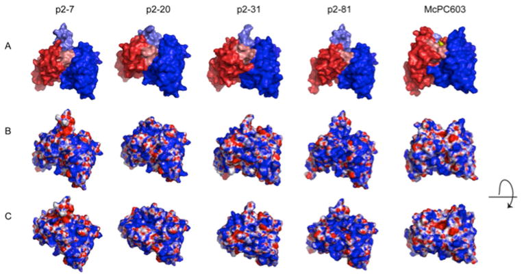
Figure 2.
Structure models of the variable region of the selected human PC antibodies. Models are shown for the anti-PC clones: p2–7, encoded by a VH3-30 and Vκ3-20; p2–20, encoded by a VH3-33 and Vλ1-44; p2–31, encoded by a VH1-2 and Vλ1-47, and p2–81, encoded by VH3-30 and Vκ1-5 rearrangements. The models are compared to the crystal structure of the PC-binding S107.1-encoded murine antibody McPC603, with the PC antigen in the binding pocket (PDB ID: 2MCP). (A) The VL region is visualized in red and the VH region in blue. HCDR3 is highlighted in lighter blue shade and LCDR3 in lighter red shade. (B) Electrostatic surface models of the variable regions. Blue color represents positively charged surface residues, red negatively charged residues. (C) Electrostatic surface models with a view looking into the potential antigen-binding site. Taken from Ref. 66.

