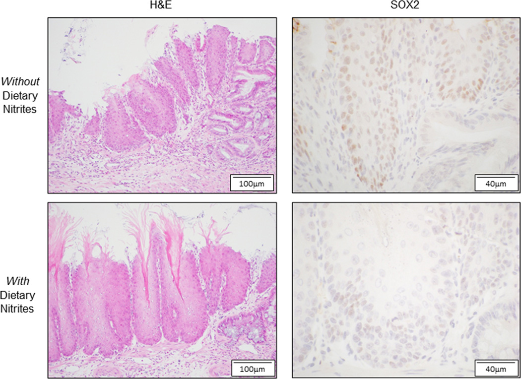
Figure 3.
In a rat model of reflux oesophagitis, dietary NO supplementation reduces Sox2 expression in the distal squamous oesophagus. Representative photomicrographs of the distal squamous oesophagus of rats with surgically-induced reflux oesophagitis that were fed diets with (lower panels) or without (upper panels) NO supplementation at 4 weeks after surgery. Inflammatory changes are seen by H&E staining in both groups. Sox2 staining is seen predominantly in the nuclei of basal and suprabasal cells, and the intensity of Sox2 staining is weaker in the rats fed an NO-supplemented diet.

