Figure 1.
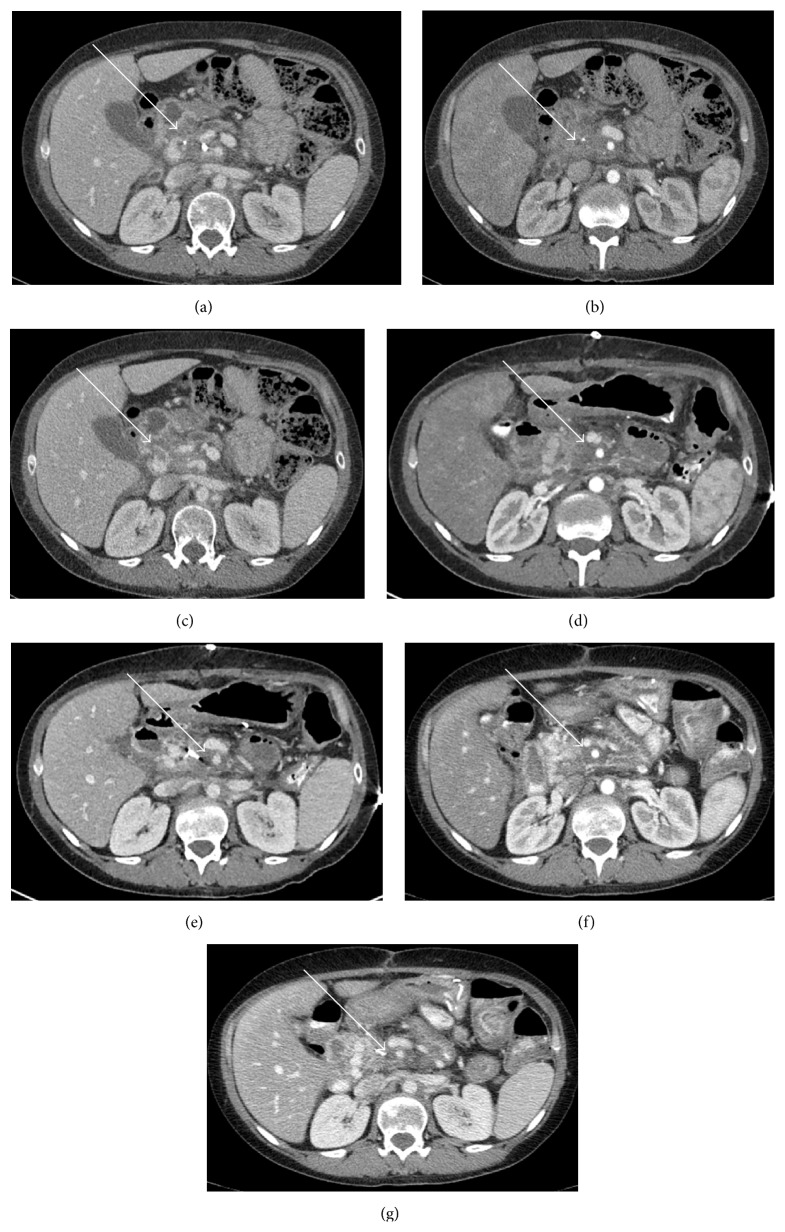
(a) Preoperative CT scan in the portal venous phase displaying abnormal soft tissue in the inferior pancreatic head (white arrow) with a nearby fiducial marker. (b) Arterial phase image displaying soft tissue encasement (white arrow) of superior mesenteric artery (SMA). Another preoperative scan (c) displaying soft tissue encasement along splenic vein and posterior to the portal confluence (white arrow). Immediate postoperative scans in arterial (d) and venous phases (e) displaying an increase in hazy soft tissue in the postablation bed (white arrows) and continued encirclement of SMA. A 2-month postoperative scan in arterial (f) and venous (g) phases displaying similar irregular, hazy, amorphous soft tissue stranding but with decreased size of the postablation zone (white arrow) consistent with no recurrence.

