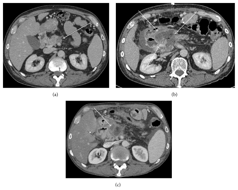
Figure 3.
(a) Preoperative CT scan demonstrating soft tissue in the posterior aspect of the proximal pancreatic body (white arrow) extending posteriorly to surround at least 180 degrees of the proximal to midsuperior mesenteric artery. (b) Immediate postoperative CT scan demonstrating a large fluid collection in ablative bed with foci of internal gas. Ablative zone (white arrows) has been demarcated from nearby duodenum (D). (c) Three-month follow-up scan showing markedly decreased ablative zone with decreased size of the fluid collection (white arrow) consistent with no recurrent disease.

