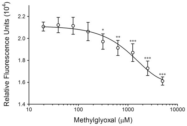
Figure 2. Prodan is displaced from drug site I by MG modification of HSA.
Prodan bound to HSA (prodan-HSA complex, 75 μM) was treated with a dilution of MG and the fluorescence (excitation 380 nm, emission 465 nm, filter cut-off 420 nm) was measured after 30 minutes. Means ± SD for three separate experiments are given. Significant (*p<0.05; ** p<0.01; *** p<0.001) as compared to control (HSA without MG treatment)

