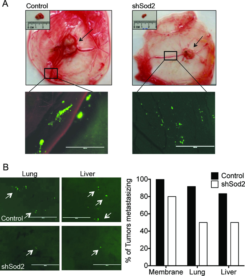
Figure 4.
Metastatic spread of ES-2 cells is attenuated with reduced Sod2 expression in the ex ovo CAM tumor model. A, Representative images show the location of the primary tumors and micro-metastasis of GFP-labeled cells in the membrane (scale bar = 1mm, CAM tumor study carried out as in Fig. 2). B, Reduced Sod2 expression inhibited tumor metastasis into the chick embryo and CAM (shSod2_#1). Quantification of percentage metastasis of tumor cells into CAM, liver and lung of chick embryo. Representative images show the percent of tumors with metastatic cancer cells present in the chicken embryo liver and lung (control, n=12; shSod2, n=10).

