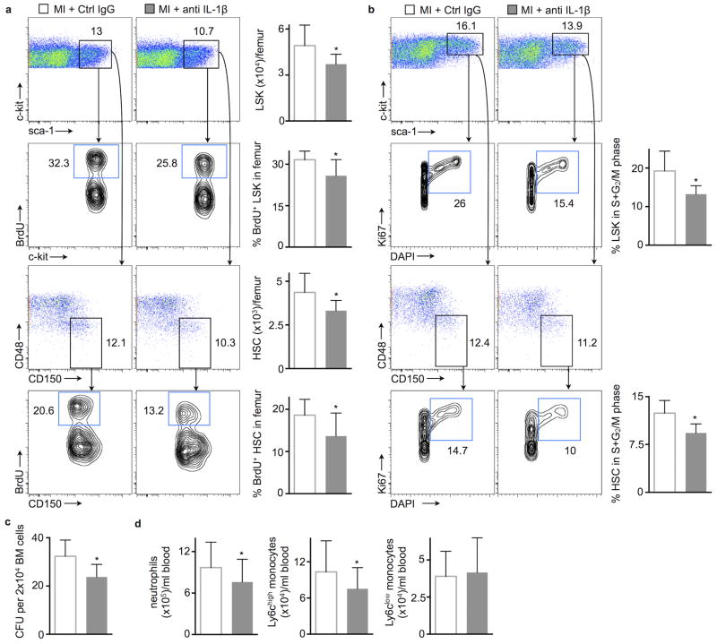
Figure 3.
Neutralizing IL-1β lowers HSC proliferation after MI. (a) Flow cytometric gating and quantification of proliferation rates with BrdU incorporation in bone marrow LSK and HSC, 48h after MI. Numbers next to gates indicate population frequencies (%) (n = 10–11 per group). (b) Cell cycle analysis in bone marrow LSK and HSC 48h after MI (n = 5–6 per group). (c) Bone marrow colony forming unit (CFU) assay 48h after MI (n = 6 per group). (d) Blood neutrophils and monocytes 7 days after MI (n = 14 per group, mean ± SD, *p<0.05, Mann-Whitney test).

