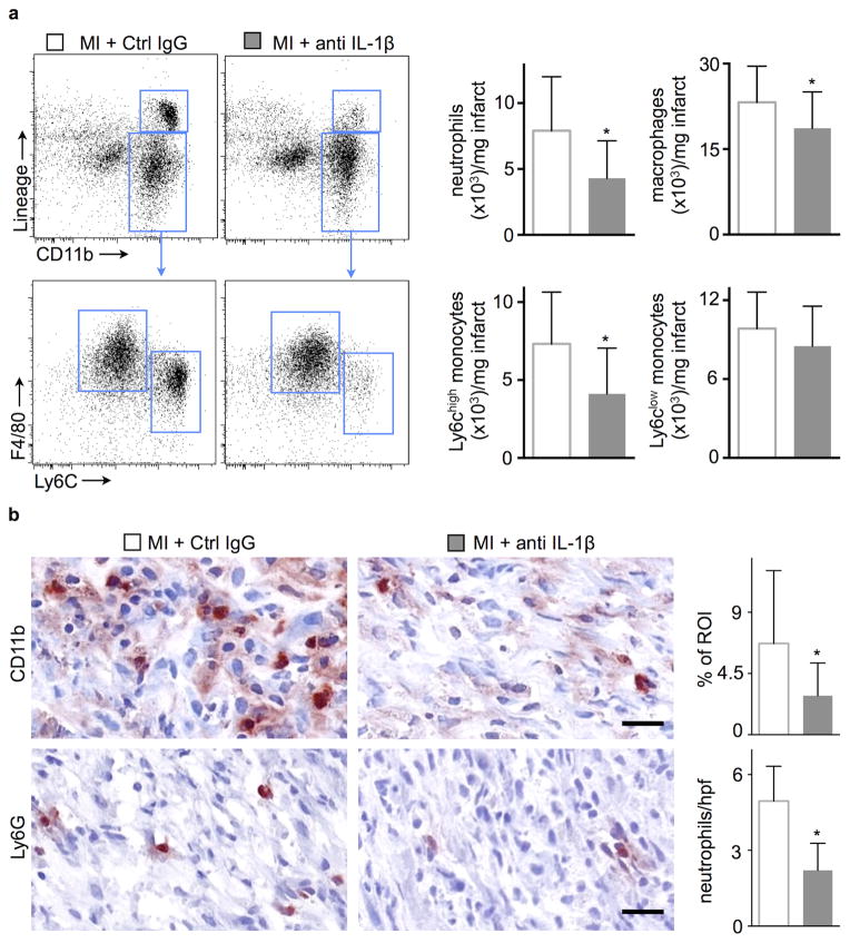
Figure 5.
Neutralizing IL-1β attenuates inflammation in infarcted hearts. (a) Flow cytometric gating and quantification of neutrophils, macrophages and monocytes in infarcted hearts 7d after coronary ligation (n=15 per group). (b) Immunohistochemical evaluation of 7d old infarcts for myeloid cells (CD11b) and neutrophils (Ly6G). Bar graphs show percentage of positive staining per region of interest (ROI) or number of cells per high power field (hpf). Scale bar indicates 50 μm (n = 6 per group, mean ± SD, *p<0.05, **p<0.01, Mann-Whitney test).

