
Figure 2.
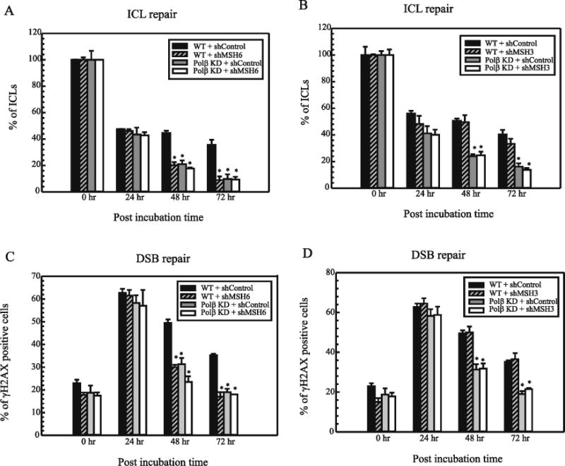
Repair of cisplatin ICLs in MDA-MB-231 cells. (A) MSH6 KD (B) MSH3 KD in MDA-MB-231. Cells were treated with cisplatin for 2 hrs and comet assays were performed as described in Materials and Methods at different time intervals (0, 24, 48 and 72 hr) to assess ICL levels. The data was collected using komet 5.5 software. The percentage of ICLs present at each time point was calculated using olive tail moments. Results are represented as mean ± SD of three independent experiments. Statistical analysis was performed by student’s t test and comparisons are made between wildtype and proficient cells vs deficient cells. NS – non-significant; * - P< 0.01. Repair of cisplatin ICL induced DSBs in MDA-MB-231 (C) MSH6 KD (D) MSH3 KD. Cells were treated with cisplatin for 2 hrs and immunofluorescence was performed as described in Materials and Methods at different time intervals (0, 24, 48 and 72 hr). A minimum of 200 cells were analyzed for each time point. The percentage of γH2AX foci positive cells at each time point was calculated. Results are represented as mean ± SD of three independent experiments. Statistical analysis was performed by student’s t test and comparisons are made between wildtype and proficient cells vs deficientcells. NS–nonsignificant;*-P<0.05.