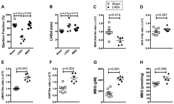
Figure 3. Elevated MBG levels contribute to nitrative stress.
Echocardiographic measures of cardiac ejection fraction (A) and Diastolic Left Ventricular Internal Dimension (LVIDd) (B) after 4 weeks of either LAD ligation (LADx) or MBG infusion. Gene expression of calcium handling proteins sarcoplasmic reticulum calcium ATPase (SERCA2a) (C) and sodium calcium exchanger (NCX-1) (D), and hypertrophic markers beta myosin heavy chain (βMHC) (E) and atrial natriuretic peptide (ANP) (F) after 4 weeks of LADx. Plasma MBG (G) levels are increased 4 weeks after LAD ligation in a post myocardial infarction heart failure model. (H) Adrenal tissue MBG levels 1 week after LAD ligation. P values were calculated using the Mann-Whitney U test.

