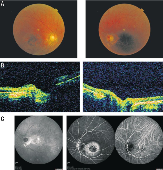
Figure 2. Ophthalmic examination results of the proband (III-12).

A: Fundus photographs of the proband showed diffuse hypopigmentation and reduced sensitivity in a central scotoma accompanied by maculopathy. The peripheral retina exhibited bone spicule-like hyperpigmentation with attenuation of retinal arteries. B: OCT of the proband revealed retinal thinning in the macular region. C: Fundus autofluorescence of the proband showed hyperfluorescence in the macula without peripheral choroidal atrophy.
