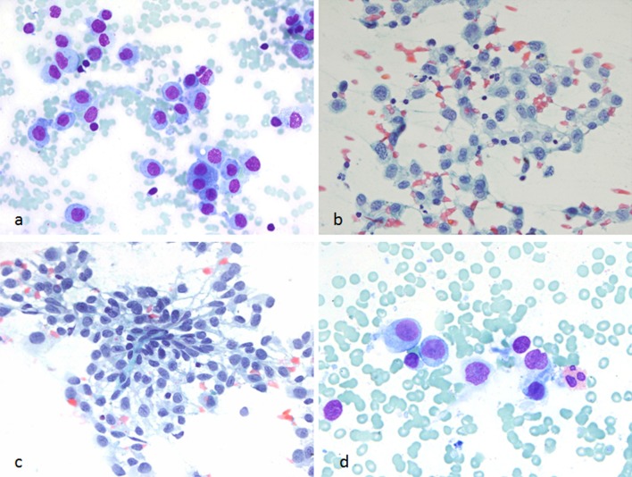
Fig. 2.
FNA smears of the thyroid mass. a Singly scattered atypical mononuclear cells seen during on-site evaluation of the aspirate (Diff-Quik stain, ×600). b, c Alcohol-fixed slides showing loosely cohesive clusters of Langerhans cells with moderate cytoplasm and convoluted nuclei (PAP stain, ×600). d Rare eosinophils were noted (Diff Quik stain, ×600, cropped and enlarged)

