Fig. 5.
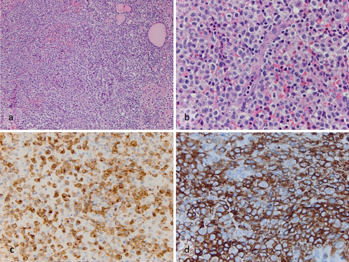
Incisional biopsy specimen from the thyroid mass. a Sheets of the neoplastic Langerhans cells and entrapped residual thyroid follicles (H&E, ×200). b Same microscopic field with higher magnification demonstrating nuclear pleomorphism of the tumor cells (H&E, ×600). c, d The diagnosis is supported by diffuse strong immunoreactivity of the tumors cells to c Langerin (Immunohistochemistry, ×400) and d CD1a (Immunohistochemistry, ×600)

