Fig. 1.
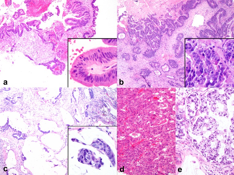
Morphologic spectrum of ITAC. a PTCC-I showing a predominantly papillary growth pattern (H&E, ×40), and stratification of fairly monomorphic nuclei resembling the appearance of a colonic adenoma (inset H&E, ×200). b PTCC-II showing a tubular and cribriform growth with stromal reaction similar to colorectal carcinoma (H&E, ×40), and more cytonuclear atypia and apoptotic debris (inset H&E, ×200). c AGC showing predominantly mucin filled spaces akin to a mucinous adenocarcinoma of colon (H&E, ×40), tumor nests are found floating in the mucin filled spaces (inset H&E, ×200). d PTCC-III showing a solid growth pattern and cytologic atypia (H&E, ×100). e Recurrence of same case showing SRC pattern instead (H&E, ×100)

