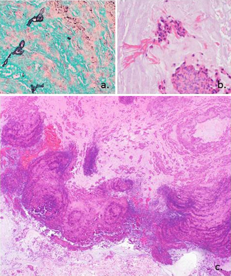
Fig. 2.
a Grocott’s methenamine silver (GMS) stain highlights the scant fungal elements seen in the specimen. b This high power image shows degenerated inflammatory cells and eosinophils with Charcot–Leyden crystals. c Hematoxylin and eosin stain shows alternating ripples of eosinophils and neutrophils creating a “tide line” or “tree ring” appearance. This low power picture is classic for allergic fungal sinusitis

