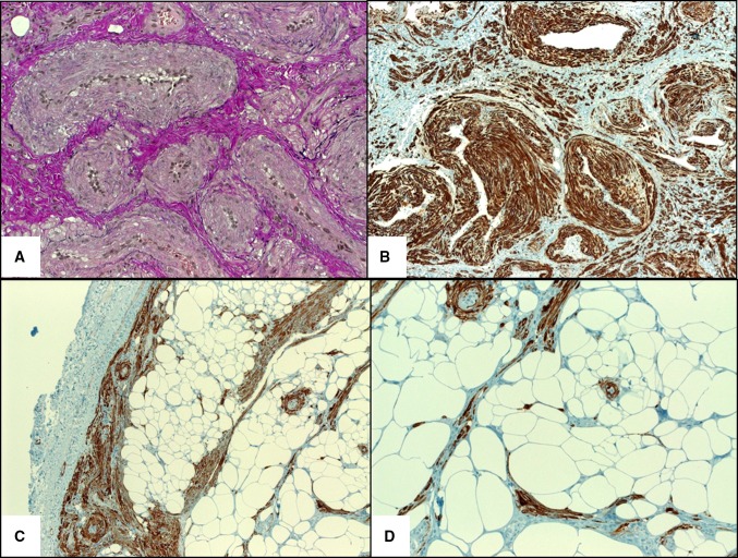
Fig. 3.
a Elastic van Gieson stain highlighting the venous channels. b Strong desmin expression highlighting intervascular smooth muscle bundles. c H-caldesmon showed strong reactivity in muscle cells (note peripheral circumscription). d Higher magnification of h-caldesmon showed scattered isolated smooth muscle cells amid the fatty component

