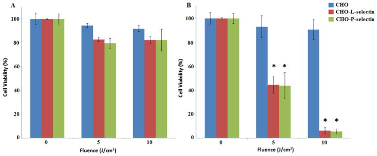
Figure 5.
PDT cytotoxicity caused by A) pyro-PEI and B) pyro-s34-PEI in CHO cells expressing P- or L- selectin. Pyro-PEI or pyro-s34-PEI was incubated with indicated cells at a concentration of 100 nM pyro in a 96-well plate and then PDT was performed using 665 nm light at the indicated fluences. Cell viability was assessed 24 hours later using the XTT assay. Mean +/− std. dev. for n=3. * denotes statistically significant difference (P < 0.05) between CHO and CHO-L-selectin or CHO-P-selectin cells based on one-way analysis of variance with post hoc Tukey’s test.

