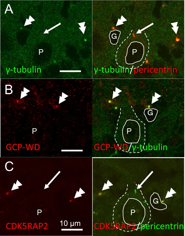
Fig. 4. .
Confocal laser microscopy for the localization of γ-tubulin and its recruiting proteins in the Purkinje cell layer of P10 cerebellar cortex. γ-Tubulin, CDK5RAP2, GCP-WD and pericentrin were co-localized at the centrosome of glial cells (double arrowheads), but γ-tubulin, GCP-WD and CDK5RAP2 were not detected at the pericentrin-positive centrosomes (arrows) of Purkinje neurons. Cell nuclei are outlined with solid lines and cell bodies are outlined with dashed lines. Nuclei of Purkinje neurons are labeled with P, and those of glia are with G.

