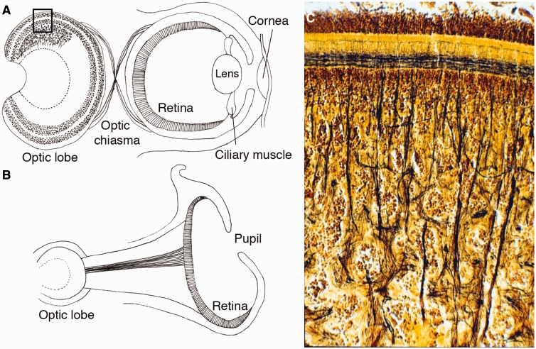
Fig. 1.
Schematic views of camera eyes and pinhole eyes of cephalopods and a photograph of a cephalopod’s optic lobe. (A) Schematic view of the cephalopod camera eye and visual system. The cephalopod retina has a single layer containing rhabdomeric photoreceptors, supporting glia, and small-caliber blood vessels in contrast to the vertebrates’ layered retina with numerous types of neurons. The visual neurons of cephalopods are located outside of the retina. The optic lobes have evolved as layered structures used in processing primary visual information. The optic lobe is the largest lobe of the brain in the all cephalopods and contains approximately 128,940,000 neurons in the brain of Octopus. The organization of the cortical layer superficially resembles that of the deeper layer of the vertebrate retina. (B) The eye of Nautilus is a unique example of a pinhole eye, which lacks lens, cornea, and contractile muscles. Nautilus’ optic lobes are simpler than those of Octopus and lack a granular cell layer. The visual neurons of octopuses and squids have a chiasma between the eye and optic lobe, but a chiasma is absent in Nautilus. (C) A cross-section (Golgi staining) showing a part of the optic lobes of Octopus corresponding to the rectangle region in A. (This figure is available in black and white in print and in color at Integrative and Comparative Biology online.)

