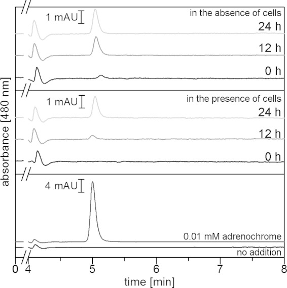
FIG 7.

Adrenochrome formation in minimal medium in the absence or presence of V. cholerae. Adrenochrome was separated by HPLC and quantified from the area of the peak eluting at 5.0 min. In the top panel (medium with 0.1 mM E in the absence of cells) and middle panel (medium with 0.1 mM E in the presence of cells), aliquots were taken at 0 h, 12 h, or 24 h and subjected to HPLC. In the bottom panel, the cell-free supernatant of V. cholerae cells was grown to the stationary phase in minimal medium without added E. Cell-free supernatant, black trace; cell-free supernatant with 0.01 mM adrenochrome added immediately before analysis, dark-gray trace. mAU, arbitrary units (103).
