FIGURE 2.
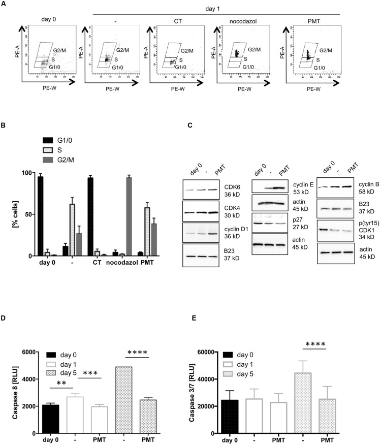
PMT stimulates cell cycle progression. (A) CD4-positive T cells were cultured overnight without activation to synchronize cells in G0 phase. Then cells were CD3/CD28-activated and stimulated with PMT for 24 h. As controls, cells were treated with 100 ng/ml CT (G0/1 phase) or 100 ng/ml nocodazole (G2 phase). Nuclei were stained with propidium iodide (PI) and intensity of the PI signal was quantified by flow cytometry (x-axis: PE-A, y-axis: PE-W). (B) Quantification of the percentage of cells in G1/0, S and G2/M phase (mean ± SD, n = 3). (C) Cell lysates were used for western blot analysis with specific antibodies against cyclin B, pCDC2 (CDK1), cyclin D1, cyclin E, CDK4, or p27. Equal loading was verified by detection of actin (whole cell lysates) or B23 (nuclear extracts). The results were corroborated with two more donors. (D,E) T cells were stimulated with PMT for 1, 3, or 5 days, lysed and the activation of caspase-8 (D) or caspases 3 and 7 (E) was measured with a Caspase-8Glo® Assay or a Caspase 3/7-Glo® Assay, respectively (Promega). Shown is the mean ± SD, n = 3 donors. Statistical analysis for this figure (B,D,E) was performed using two-sided ANOVA for multiple comparisons (∗∗p ≤ 0.01; ∗∗∗p ≤ 0.001; ∗∗∗∗p ≤ 0.0001).

