FIGURE 3.
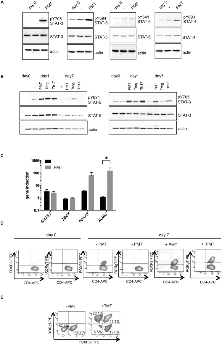
PMT induces Foxp3 and RORγt-positive T cells. CD3/CD28-activated T cells were stimulated with PMT overnight or for 7 days, respectively. (A) Whole cell lysates were produced and antibodies against p(Y705)STAT-3, STAT-3, p(Y694)STAT-5, STAT-5, p(Y641)STAT-6, STAT-6, and p(Y693)STAT-4, STAT-4 were used for immunoblotting. Equal loading of protein was verified by actin detection. (B) As a positive control, T cells were treated with 100 ng/ml IL-1β, IL-23, IL-6, TGF-β (Th17 cells), or 100 ng/ml IL-2 and rapamycin (Tregs). (C) Quantitative RT-PCRs were performed using SYBR Green and sequence specific primers for FOXP3, TBET, GATA3, and RORC. Results were normalized against the housekeeping gene actin. Shown is the mean of induction compared to non-activated cells. SD, n = 2 donors, experiment performed in duplicates. Statistical analysis was performed using two-sided ANOVA for multiple comparisons (∗p ≤ 0.05). (D) Intracellular FACS analysis. Fixed and permeabilized cells were stained with anti-CD4-APC, purified anti-Foxp3 or anti-RORγt and FITC-/PE-labeled secondary antibodies. (E) Intracellular double stain of Foxp3 or RORγt of cells on day ten. Data shown in (A,C,D) are representative for three, and (E) for two experiments.

